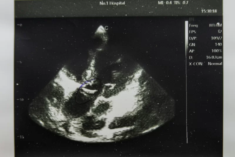
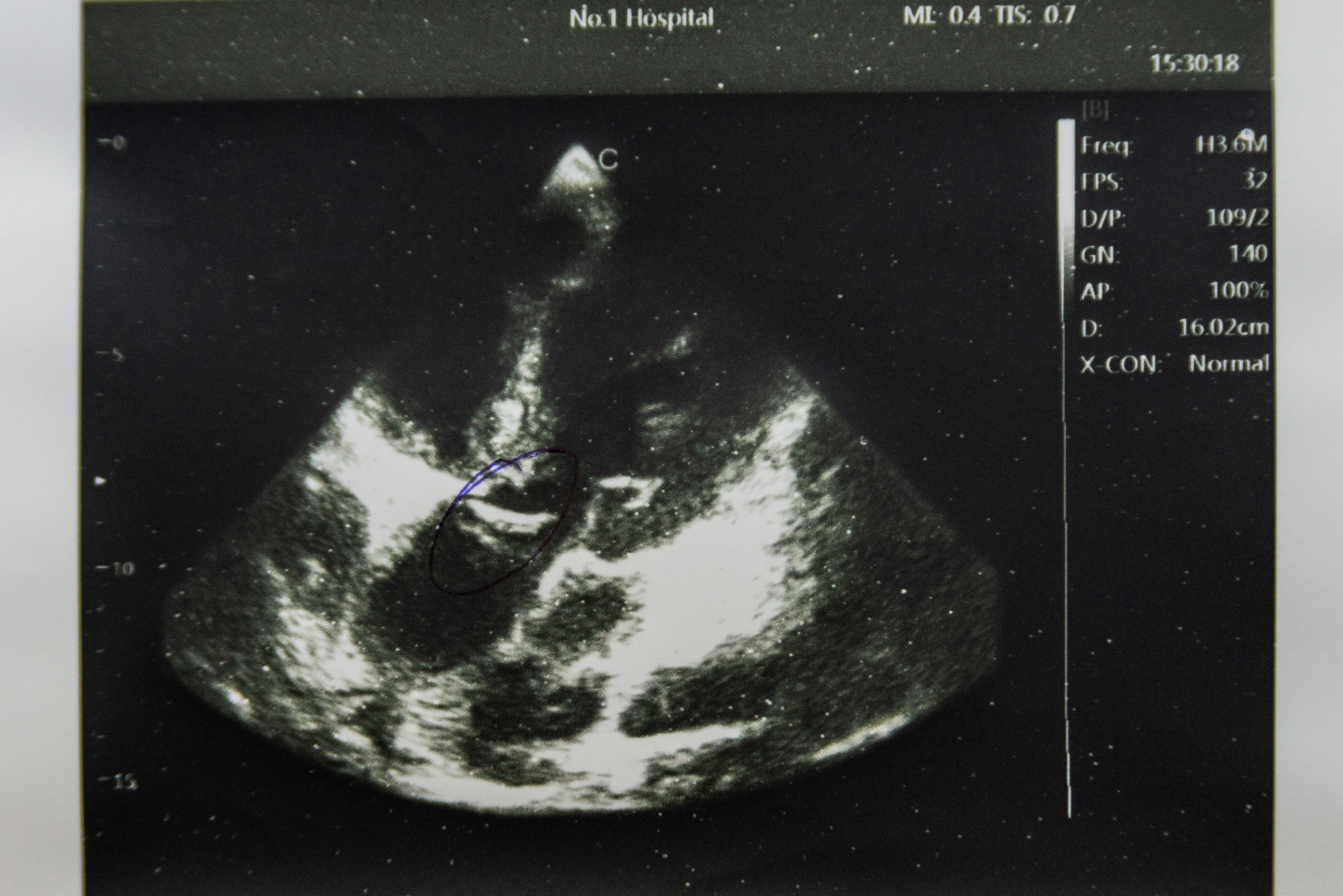
This image presents a high-quality snapshot obtained from a cardiac ultrasound examination. The structures of the heart valve are clearly visible on the screen, highlighted by an oval outline, indicating a detailed study of its condition. The depth of the examination, indicated by the scale on the left, allows for the assessment of the location and dimensions of the area under investigation. The presence of text with parameters such as 'Freq', 'FPS', 'D/P', 'GN', 'AP', 'D', and 'X-CON: Normal' signifies professional medical equipment and precise diagnostics. The examination time, 15:30:18, and the institution's name, 'No.1 Hospital', add context to the image, making it valuable for medical archives, educational purposes, or as an illustration in cardiology articles.
The black and white palette of the image is typical for ultrasound diagnostics, with different shades of gray conveying tissue and fluid density, enabling physicians to analyze blood flow and heart structure. The detail of the valve, its mobility, and integrity can be key factors in diagnosis, making such a clear image highly significant. The absence of obvious pathologies, as suggested by the 'X-CON: Normal' label, might indicate a healthy state of the examined valve, but final conclusions are exclusively made by a specialist.
This image is an excellent example of modern medical imaging, demonstrating the capabilities of ultrasound in examining internal organs. It can be used for training medical students, illustrating the functionality of cardiological equipment, or as part of a broader set of images related to health and medicine. The clarity of the image and the presence of informative markers make it a useful tool for specialists and a general audience interested in healthcare advancements.


